UPJ OBSTRUCTION
UPJ OBSTRUCTION
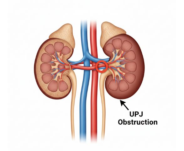
A UPJ obstruction (Ureteropelvic Junction Obstruction) is a blockage at the junction where the kidney meets the ureter, which restricts the normal flow of urine from the kidney to the bladder. This condition can be present from birth or develop later due to scarring, compression from nearby blood vessels, stones, or inflammation. When left untreated, UPJ obstruction may lead to kidney swelling (hydronephrosis), pain, recurrent urinary tract infections, or reduced kidney function.
Causes of UPJ Obstruction
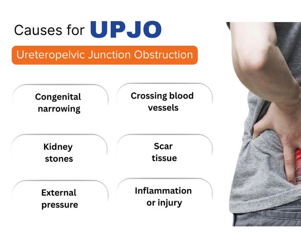
UPJ obstruction occurs when a blockage at the ureteropelvic junction prevents urine from flowing normally from the kidney to the ureter. This leads to urine buildup in the kidney (hydronephrosis) and may affect kidney function over time. The main causes include:
Congenital narrowing – Many cases are present from birth due to abnormal development of the ureter.
Crossing blood vessels – Nearby blood vessels may compress the ureter, causing obstruction.
Scar tissue or strictures – Inflammation, previous surgery, or injury can lead to narrowing at the junction.
Kidney stones – Stones can block or irritate the UPJ, leading to obstruction.
Infections or inflammation – Recurrent urinary infections may cause swelling and scarring.
Previous procedures or trauma – Surgical interventions or injury to the kidney or ureter may result in obstruction.
Symptoms of UPJ Obstruction
✔ Flank or upper abdominal pain
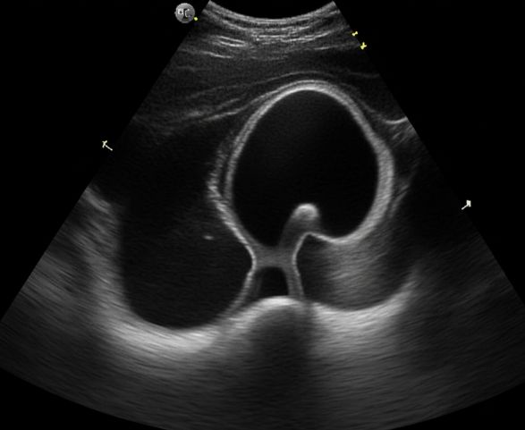
✔ Swelling of the kidney (hydronephrosis) seen on imaging
✔ Recurrent urinary tract infections (UTIs)
✔ Blood in the urine (hematuria)
✔ Nausea or vomiting during pain episodes
✔ Fever in cases of infection
✔ Reduced urine output in severe cases
✔ Poor growth or feeding difficulties in infants and children
Diagnosis of UPJ Obstruction
🔹 Ultrasound – First-line imaging to detect kidney swelling (hydronephrosis) and assess urine buildup.
🔹 CT Scan or MRI Urography – Provides detailed images of the kidneys and ureter to identify the exact site and cause of obstruction.
🔹 Diuretic Renal Scan (MAG3/DTPA) – Evaluates kidney function and measures how well urine drains from the kidney.
🔹 Intravenous Pyelogram (IVP) – An X-ray with contrast dye to visualize urine flow from the kidneys to the bladder.
Treatment Options for UPJ Obstruction
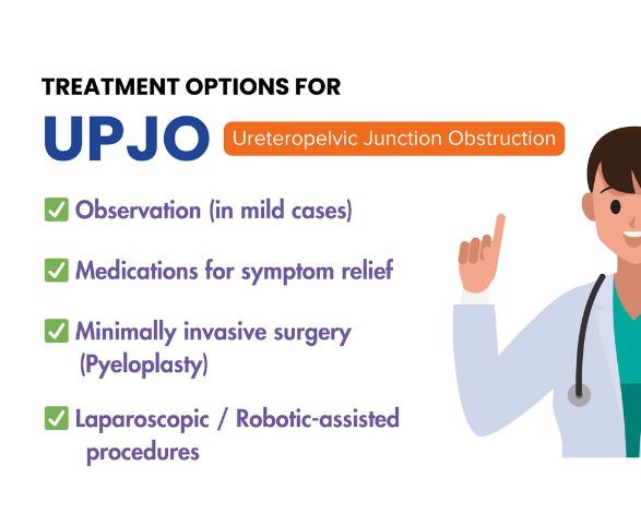
✅ Observation & Monitoring – Mild or asymptomatic cases may be managed with regular follow-up scans to monitor kidney function and drainage.
✅ Endopyelotomy – A minimally invasive endoscopic procedure where the narrowed junction is incised to improve urine flow.
✅ Pyeloplasty (Laparoscopic/Robotic/Open) – The gold-standard surgical treatment that removes the blocked segment and reconstructs the ureter for long-term relief.
✅ Temporary Stenting or Nephrostomy – Used to immediately relieve obstruction and drain urine while preparing for definitive treatment.
✅ Medical Management – Antibiotics and pain control may be used to treat infections or symptoms associated with UPJ obstruction.
Why Choose Shivasya ?
✔️ Advanced Minimally Invasive & Laser Procedures
✔️ High Success Rates with Urethroplasty
✔️ Experienced Urologists
✔️ Specialized Urological Care
✔️ Comprehensive Post-Treatment
✔️ Follow-Up and Support
📅 Book Your Appointment Today for expert evaluation and treatment of urethral stricture.
💙 Your comfort, safety, and recovery are our top priorities at Shivasya Urology Hospital.
🏥 Trusted care, advanced technology, and compassionate healing — all under one roof.
PLAN A VISIT TO YOUR DOCTOR
HOSPITAL ADDRESS
4th Floor (401, 402), Shrikunj ,
Icon, 1/A Vitthal Bhai Patel Colony,
Nr. Lakhudi Cross Road,
Navanrangpura, Ahmedabad,
Gujarat – 380 009, India.
+91 79 2991 6245
+91 99258 89372
shivasyahospital@gmail.com